| . |
| Scrutinizing the evidence for breast cancer procedures and treatments |
| Imaging FAQ |
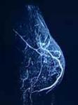
How should I be screened for breast cancer? In addition to physician exams and breast self-examination, there are primarily three breast imaging techniques: mammograms (X-rays), ultrasounds, and MRIs (with contrast dye). A newer technique, thermography, is still being studied. In his chapter, "The Danger and Unreliability of Mammography: Breast Examination is a Safe, Effective, and Practical Alternative," (The International Journal of Health Services,Volume 31, Number 3 [2001] ), Dr. Epstein states, "Mammography screening is a profit-driven technology posing risks compounded by unreliability. In striking contrast, annual clinical breast examination (CBE) by a trained health professional, together with monthly breast self-examination (BSE), is safe, at least as effective, and low in cost." He goes on to say, "Mammography is not a technique for early diagnosis. In fact, a breast cancer has usually been present for about eight years before it can finally be detected. Furthermore, screening should be recognized as damage control, rather than misleadingly as secondary protection." (See Epstein, S., International Journal of Health Services 2001, in the MEDICAL ARTICLES' IMAGING section, or go to: Dr. Epstein on Mammography. Please consider the data in this section of the FAQ comparing mammograms, ultrasounds, MRIs and thermograms before rushing into any single diagnostic procedure. PRINT OUT THE ARTICLES FOR YOUR PATIENT PORTFOLIO. Is there much radiation in mammograms? UPDATE: In a March 2006 study, low energy X-rays used in mammograms were found to cause approximately four times, but possibly as much as six times, more mutation damage than higher energy X-rays. Since radiation risk estimates are based on the effects of high energy radiation, this implies that the risks of radiation- induced breast cancers for mammograms are underestimated by the same factor. (See Heyes GH et al., Enhanced Biological Effectiveness of Low Energy X-rays and Implications for the UK Breast Screening Programme, Br J Radio 2006.) Mammography Consideration: For Women Between 40 and 49 A 2006 study suggests that surgical removal of breast tumors in young women may instigate angiogenesis (new vessel growth) by possibly removing inhibitors of angiogenesis or promoting growth factors in response to wounding in dormant distant disease in approximately 20% of cases of premenopausal node-positive women who have not had chemotherapy. Women need to be advised of the risk of accelerated tumor growth and early relapse before giving informed consent to mammography. (See Retsky M et al., Does Surgery Induce Angiogenesis in Breast Cancer? Indirect Evidence from Relapse Pattern and Mammography Paradox Int J Surg 2005. Plus, see correspondence in New England Journal of Medicine, February 16, 2006.) How reliable are mammograms in detecting breast cancer? Although new mammography technology has increased the accuracy of detecting breast cancer, it still remains more of a challenge to mammographically detect breast cancer in women with non-fatty, dense breasts (common in women under 50). Has the advent of computer-aided mammograms helped to detect breast cancer in women with dense breasts? A June 2006 retrospective study of malignant tumors found better detection using computer-aided mammograms dependent upon breast density types. For example, with lesser breast density, type 1, there was an 84.85% accuracy rate in detecting malignancy, while for dense type 4 breast tissue, there was a 69.70% accuracy rate. (See Obenauer S et al., Impact of Breast Density on Computer-Aided Detection in Full-Field Digital Mammography, J Digit Imaging 2006.). If I have a lump in my breast and it turns out to be cancer, can the mammogram spread it by compression ? Dr. Robert Rosser, in his article, "Point of View: Trauma is The Cause Of Occult Micrometastatic Breast Cancer in Sentinel Axillary Lymph Nodes," published in the medical journal, The Breast (2000), cautions against compressing, squeezing, or otherwise disrupting tumors that may result in trauma-induced micrometastases. He calls these dislodged cancer cells "traumets." Especially if a lump is known to be present at the time of the mammogram, Dr. Rosser advises that the compression of the breast should be minimal to prevent compression-induced traumets. While the risk of traumets may be small, if a patient hasn't yet had a diagnostic mammogram, other non-compressing imaging procedures may be considered. Dr. Rosser suggests that the time between the mammogram and the surgical excision of the traumet may determine whether an insignficant traumet will divide, grow and progress to a real metastasis that may spread from the lymph nodes and elsewhere if not removed. (See Rosser, RJ, The Breast, The Breast 2000, in the MEDICAL ARTICLES' IMAGING section.) Other Imaging Procedures to investigate: Ultasound and MRI How does an ultrasound compare to a mammogram and an MRI ? Ultrasound (also called a sonogram), which does not use compression, radiation, or dye, is generally useful for screening women with dense breasts. The MRI, which uses a contrast dye, is similarly useful in women with dense breasts. In a study assessing the accuracy of mammography, clinical examination, ultrasonography, and magnetic resonance (MRI) imaging in the preoperative assessment of the local extent of the breast cancer, ultrasound showed higher sensitivity than mammograms for invasive ductal carcinoma in nonfatty breasts. (See Berg WA et al., Diagnostic Accuracy of Mammography, Clinical Examination, US, and MR Imaging in Preoperative Assessment of Breast Cancer, Radiology 2004.) What is a Breast MRI? Please go to this link for a full explanation. What is a Breast MRI? What if I feel a lump? Update: In a May-June 2006 study, ultrasounds were found to be more accurate in measuring palpable tumors (tumors that can be felt) than clinical exams or mammograms. As determined by pathological exam, the maximal tumor diameter was within 2mm of the pathologic tumor size in 45.2% of ultrasounds, 28.2% of mammograms, and 14.5% of clinical measurements. (See Shoma A et al., Ultrasounds for Accurate Measurements of Invasive Breast Cancer Tumor Size, Breast J 2006.) In addition, in another 2006 study, axillary ultrasonography was found to be helpful in differentiating metastatic lymph nodes from normal lymph nodes. (See Wang YB et al., Evaluation of the Ultrasonographic Features of Axillary Lymph Node Metastasis in Breast Cancer, Clinical Oncology Institute, 2006.) In the case of a palpable tumor, a mammogram is given in tandem with an ultrasound, and many doctors and breast centers in the US resist doing an ultrasound (no compression and no radiation) alone. Often, only persistence on the patient's part will allow the patient to undergo an ultrasound alone. A recent German study, cited below, suggests that ultrasound may be the "preferred imaging procedure" for a palpable tumor ( a tumor which can be felt.) During an ultrasound, the breast isn't compressed or exposed to radiation. Usually, a mammogram is given in tandem with an ultrasound, and many doctors and breast centers in the US resist doing an ultrasound alone. Often, only persistence on the patient's part will allow the patient to undergo an ultrasound alone. -----------------------------------------------------------------
Re-evaluating the role of breast ultrasound in current diagnostics of malignant breast lesions Hille H, Vetter M, Hackeloer BJ. Praxis fur Gynakologie und Geburtshilfe, Hamburg. CONCLUSIONS: Breast ultrasound should be the preferred imaging procedure in the case of a palpable lump, leading to a definitive diagnosis itself or with an additional consecutive core needle biopsy. For women without symptoms, breast sonography should be mandatory and complementary to mammography in the case of breast density grade II (BI-RADS) or more.
(See more literature on mammograms, ultrasound and MRI in the MEDICAL ARTICLES' IMAGING section.) |
. |
Take home questions to ask yourself: 1. If other reliable imaging procedures are available, do you want to risk having a mammogram if the compression during the mammogram is more than minimal? 2. If you've already had a mammogram, can you shorten the time between the mammogram and surgery date to minimize the chance of traumets before they can become real "mets" (metastases)? |


Join our email list for only. Your information will not be shared or sold to anyone. |
1. If other reliable imaging procedures are available, should do you want to risk having a mammogram if the compression during the mammogram is more than minimal? 2. If you've already had a mammogram, can you shorten the time between the mammogram and surgery date to minimize the chance of traumets before they can become real "mets" (metastases)? |
When weighing the safety of all three mainstream procedures (mammography, MRI and ultrasound), we must consider the radiation toxicity of the mammogram and the chemical toxicity of the contrast dye in the MRI procedure. Of the three mainstream used more widely but are still being evaluated for reliability. Each patient needs to study each method before making an informed decision. |
Breast Cancer ChoicesTM

| Read The Lancet Oncology on screening mammography |
Do 35% of invasive cancers spontaneously regress once they reach 1 cm? |
- These statements have not been evaluated by the U.S. Food & Drug Administration. The
information discussed is not intended to diagnose, treat, cure, or prevent any disease.
- This website is intended as information only. The editors of this site are not medically-trained.
Please consult your licensed health care practitioner before implementing any health strategy.
The information provided on this site is designed to support, not replace, the relationship that
exists between a patient/site visitor and his/her existing physician. This site accepts no
advertising. The contents of this site are copyrighted 2004-2015 by Breast Cancer Choices,
Inc., a 501 (c) (3) nonprofit organization run entirely by unpaid volunteers.
Contact us with comments or for reprint permission at admin@breastcancerchoices.org
| Home FAQ Strategies Discussion Iodine Hormones Contact Store |